Processing pipeline details
fcdproc is comprised of 3 levels: Preprocessing, Modeling, and Detection
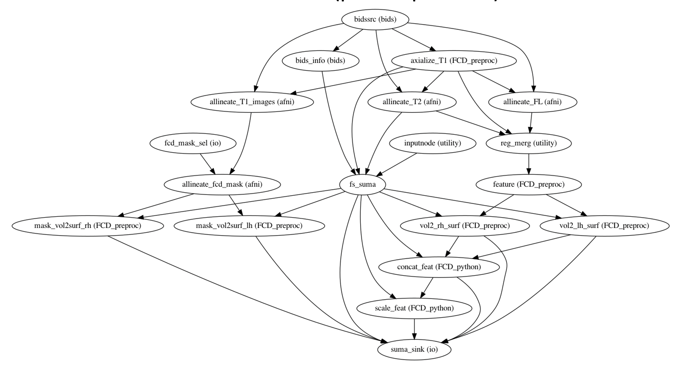
Preprocessing of structural MRI
The anatomical sub-workflow expects 3 MRI contrasts as its inputs (T1, T2, and FALIR). then it performs the follwoing steps:
T1 axialization with respect to TT_N27 template
placed under $bids_dir/__files/TT_N27.niiT2 and FLAIR coregistration to T1
subject specific intensity normalization
feature generation
surface reconstruction (fs-suma)-details in the follwoing section
surface feature normalization

Note
for FCD positive patients who have their fcd_mask dervied from the original input T1, please place them under $bids_dir/mask/$subject_id/fcd.msk.nii. The single subject workflow would then coregisters the input T1 to the axialized T1, apply the transformation metrix to the fcd.msk.nii to get it in alignment with T1 and then maps them from volume domain to surface domain.
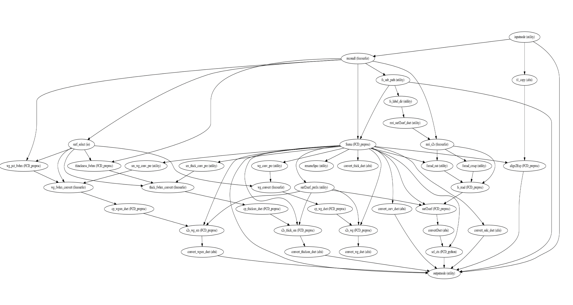
The fs-suma workflow perform freesurfer’s reconall to reconstruct the brain surface from T1 and T2 structural images.
If enabled, several steps in the fcdproc pipeline are added or replaced.
All surface reconstruction may be disabled with the --fs_no_reconall flag.
In order to bypass reconstruction in fcdproc, place existing reconstructed
subjects in <output dir>/freesurfer prior to the run, or specify an external
subjects directory with the --fs_subjects_dir flag.
fcdproc will perform any missing recon-all steps, and continue with SUMA to convert
the cortical surface into AFNI-based fromat.

Normative Modeling
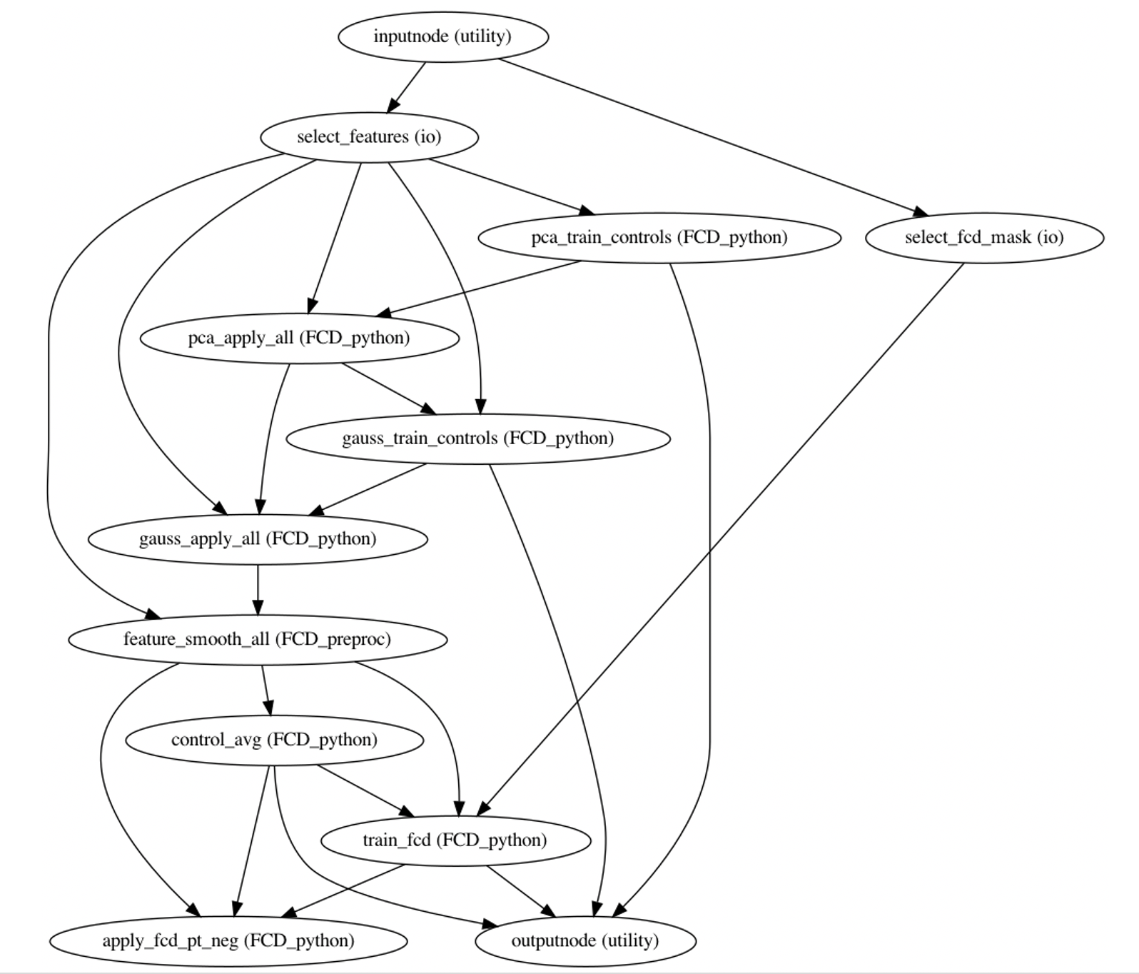
We perfrom the following steps to train our FCD-detector model:
dimensionality reduction of the initial feature vectors across HVs to components required to explain 90% of the variance
PCAiterative non-linear gaussianization to obtain a multivariate normal distribution of features
GAUSSsurface-based smoothing and averaging of features across HVs at each vertex on the gray-white junction surface
control_avgThe same features were computed at each vertex within the training FCD masks in MRI+ patients to generate a normalized average FCD feature vector
fcd_detector

FCD Lesion Detection
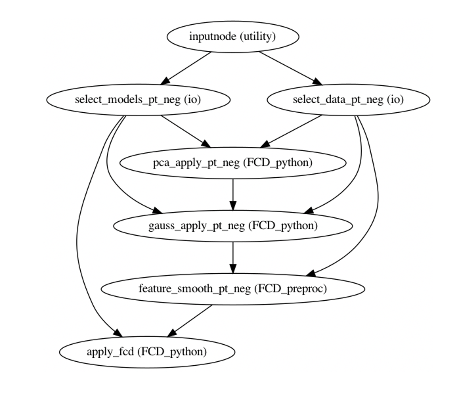
At this step, smoothed normalized features at each vertex were projected onto the FCD average unit vector, allowing for estimation of similarity to FCDs, then corrected for the expected appearance at each vertex.
